I recently worked on a model of a 3D disarticulated skull with Pixologics ZBrush. It was quite a large project to take on in the way of ensuring accuracy and especially the accuracy of the sutures in order for a perfect fit, one piece to the next. This is a temporary turntable that needs updating with the many parts of the ethmoid remaining together as one piece. I will reveal the production process involved in this later in the year. In the mean time if you want to see it in high quality print you should check out Dorling Kindersleys 'The Complete Human Body' which I worked on heavily last year and earlier this year.
Thursday 2 September 2010
Wednesday 26 May 2010
Disarticulated Skull
Well its been a while since I posted anything on here as time really hasn't permitted as i have had lots on the go. I recently received my disarticulated skull from Bone Clones, which cost me in all around £1050, itha tax and postage etc. I have produced a very detailed 3D disarticulated skull which will appear in a book to be released in September, but more about that at a later date. In the mean time here is something I came across in my research which in my opinion is one of the better 3D disarticulated skulls out there, along with a very useful turntable animation and dialogue.
Friday 26 February 2010
Sunday 21 February 2010
La Specola
Long before Plastination changed the way the public were able to view anatomy there was a trend for making anatomical replicas in wax between the late 1500's and the 1700's. There is a large collection of these wax anatomical specimens on display at the Museum of Zoology and Natural History "La Specola" Florence, within 10 of its 34 exhibiting rooms.
Some amazing photos on Flickr
extract from the museums website:
The Museum is particularly proud of its collection of anatomic waxes, an art introduced in Florence by Ludovico Cigoli (1559-1613), which enjoyed its maximum period of splendour and technical and scientific accuracy during the 18th century. The most famous representative of wax sculpture was Clemente Susini (1754-1814) who made the most important pieces of the collection in the laboratory of the Museum (that has not been in use for Down a century).
The most important pieces of the wax collection is represented by the group of waxes by Gaetano Zumbo (1656-1701), which possess an extraordinary artistic value besides representing excellent anatomical models.
The most important pieces of the wax collection is represented by the group of waxes by Gaetano Zumbo (1656-1701), which possess an extraordinary artistic value besides representing excellent anatomical models.


Saturday 20 February 2010
Wellcome Museum of Anatomy and Pathology
Whilst there are traveling exhibitions displaying human anatomy in a way we may presume has never been done before there are in fact many institutes and museums offering a much greater learning experience and better opportunities to come in to close contact with anatomical specimens. One which I am looking to visit in the coming month is the Wellcome Museum of Anatomy and Pathology which is designed to be a learning resource for students and professionals that study or work within the medical arena. With over 2000 wet preparations (A method of preparing specimens for examination in which they are kept in their liquid state or suspended in a liquid, rather than being dried and then examined.) , disarticulated bones, models and slides amongst many other interesting learning aids. You can visit there as part of a group study outing or even arrange for a private study and is often booked for large groups and presentations, as well as offering workshops and courses. The address is Royal College of Surgeons of England, 35-43 Lincoln's Inn Fields London. TEL: 020 7869 6560 for visiting inquiries.


BODIES on exhibition
Since Gunthar von Hagens developed and released the plastination process, and with the success of the 'BODY WORLDS' exhibitions there have been numerous similar exhibitions presenting anatomy in more or less the same manor. One of the main differences between Body Worlds and other 'body shows' such as Bodies the Exhibition is how the specimens are sourced. Many people are now aware of the volunteer system that was set up by GvH and co. and that they no longer need to purchase and source bodies from certain controversial suppliers as previously covered by the international media. Although it is apparent that the specimens at other exhibitions have been sourced in not such an ethical manner.
Looking at the skeletal structure of the specimens on show at Bodies the Exhibition in New York I noticed extremely thin bones of the pelvis and scapular of many of the bodies on show, which from what I understand is a clear sign of illness or malnutrition. One of the specimens I looked at even had perforations through the Ilium. I did quite a lot of research into this subject for my dissertation and was quite surprised at what I discovered.
On a positive note one of the most amazing things that I saw was within a darkened room filled with many glass presentation cabinets containing whole or sectioned parts of the cardiovascular system, with the arteries dyed in red. The main lighting in the room was from within the cabinets which brilliantly illuminated the specimens. Here is an example from Body Worlds 4:


 All images Copyright ©Gunther von Hagens, Institute for Plastination, Heidelberg, Germany,
Extract from Bodyworlds web site:
Dr. Gunther von Hagens is the world’s leading anatomist, the inventor of Plastination (the method of specimen preservation that makes anatomical display possible), and the originator of BODY WORLDSKOERPERWELTEN (its German title), the first of its kind anatomical exhibition. Dr. von Hagens invented Plastination at the University of Heidelberg in 1977, developed the use of plastinated specimens for medical education and preservation, and established the International Society for Plastination. More than 400 universities in 40 countries utilize Dr. Gunther von Hagens’ preservation technique in their curriculum.
Looking at the skeletal structure of the specimens on show at Bodies the Exhibition in New York I noticed extremely thin bones of the pelvis and scapular of many of the bodies on show, which from what I understand is a clear sign of illness or malnutrition. One of the specimens I looked at even had perforations through the Ilium. I did quite a lot of research into this subject for my dissertation and was quite surprised at what I discovered.
On a positive note one of the most amazing things that I saw was within a darkened room filled with many glass presentation cabinets containing whole or sectioned parts of the cardiovascular system, with the arteries dyed in red. The main lighting in the room was from within the cabinets which brilliantly illuminated the specimens. Here is an example from Body Worlds 4:

and here is another interesting example showing the renal arteries:

and finally the lower arm:

Extract from Bodyworlds web site:
Dr. Gunther von Hagens is the world’s leading anatomist, the inventor of Plastination (the method of specimen preservation that makes anatomical display possible), and the originator of BODY WORLDSKOERPERWELTEN (its German title), the first of its kind anatomical exhibition. Dr. von Hagens invented Plastination at the University of Heidelberg in 1977, developed the use of plastinated specimens for medical education and preservation, and established the International Society for Plastination. More than 400 universities in 40 countries utilize Dr. Gunther von Hagens’ preservation technique in their curriculum.
Sunday 14 February 2010
Maxilla & Mandible
I have a small model of a skull in front of me on my desk and for its size it holds a pretty good level of quality and has a nice feel to it. I purchased it a few years back in New York from a store called Maxilla & Mandible, located at 451 Columbus Avenue, between 81st and 82nd Streets in New York City. On my visit to NY I naturally went in search of what ever I could find in relation to Human Anatomy and this store was just one of my findings. It is like walking into a natural history museum when your inside, although of course much smaller, but with a large array of products and specimens. Its hard to tell from their web site how fascinating the store actually is. Its not only Human anatomy within, but also Paleontology, Osteology, Entomology and Malacology. Here is a video featured on the web site showing some of the publicity they have had.

And here is the little skull I purchased:
Detailing a Skull
I have been working on a large project now for around six months, and now it is in its final stages. Working with a company called Medi-Mation I have been using Zbrush to texture, edit and add to an existing commercial anatomical model. I won’t go into much detail on here as of yet until the project is complete and a bit more out in the open. One thing I can talk about is a part of the project that relates to my personal work, and that is creating a disarticulated skull. There are many digital models available on the market today and quite a few skeletal systems, and many more skulls, but only a few disarticulated skulls. Something I have been working on and will be taking further today is a 'very' disarticulated skull with even the ethmoid separated in order for the viewer to achieve further understanding. This is not a particularly easy piece of work, primarily because of what is available to research. I am using as much MRI data as I can find a complete cast of a skull, a disarticulated cast of a skull, Sobotta's Atlas and Gray's Atlas, and scanning the internet for photographs.
I have searched around for a 'museum quality replica' and there are a few, for around £800-£1000 and so it’s a big pay out in order to ensure correct detailing. The plan of action is to work in stages, and so I have decided to complete stage one as a basic model suitable for s single rendering purpose, and then do some reading in order to understand the form of the skull better before either purchasing a an mq replica or visiting universities and museums with my camera. I will post some sneak previews on here soon, but it will most probably be of a single piece, and a complete skull, leaving the full reveal until completion.
I have searched around for a 'museum quality replica' and there are a few, for around £800-£1000 and so it’s a big pay out in order to ensure correct detailing. The plan of action is to work in stages, and so I have decided to complete stage one as a basic model suitable for s single rendering purpose, and then do some reading in order to understand the form of the skull better before either purchasing a an mq replica or visiting universities and museums with my camera. I will post some sneak previews on here soon, but it will most probably be of a single piece, and a complete skull, leaving the full reveal until completion.
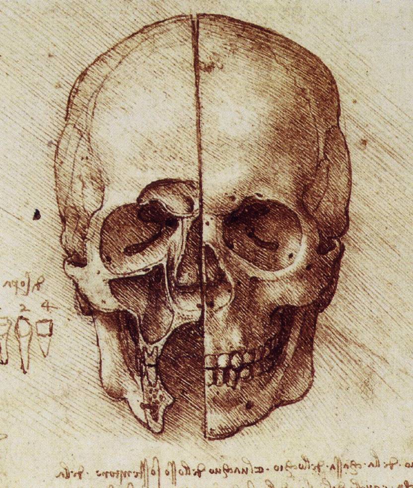
A sketch by Leonardo da Vinci
Saturday 13 February 2010
The Begining
I have finally started this blog as planned for months. After graduating and pursuing my career as a Medical/Anatomical Illustrator I have spent hour upon hour searching the internet and book shops in order to find the best reference and inspiration. This blog is for my own personal use as a way of organising all the reference material I found and for those of you who share my interest in medicine and anatomy. Of course its also a way for me to exhibit my own illustration, in progress, and in publication. So here it goes..........
Subscribe to:
Posts (Atom)

